Personal notes on the article:
This is a short article from 1950 about the episacroiliac lipomas or back mice as a cause of low-back pain written by Kermit Katz and Morton Berk from the Boston City Hospital.
The article was published in The New England Journal of Medicine. They present an illustrative case report about the fatty tumors called episacroiliac lipomas.
Surprisingly, in 1950, Katz and Berk stated in the introduction of this article:
“From recent reports in literature and from our own experience, it appears probable that this entity –episacroiliac lipomas– deserves prominence as a MAJOR CAUSE of the elusive clinical symptom of low-back pain.”
-They mention the work of Sutro (1935), Ries (1937) and Copeman and Ackerman (they made a nice summary about these authors’ work).
-They mention that in the English literature these nodules became to be called “fibrositis”.
-They summarize that the PAINFUL TENDER LOBULES OF FAT or episacroiliac lipomas occur commonly in the sacroiliac region, and had recently been recognized as a cause of low back pain (AT LEAST that was in the 50’s, afterwards they became unrecognized by probably various reasons).
-They emphasize that obesity seems to be an etiological factor, since all their patients presented in the article suffered from obesity and low-back pain with tender NODULATIONS in the sacroiliac area. They hypothesize that the fatty tissue herniates through the fascial planes.
-It is important to notice that they presented 5 cases of OBESE WOMEN that DID NOT improve with the local injection of anesthetic as other authors pointed out. The patients did a PROMPT IMPROVEMENT with the surgical removal of the fatty mass.
Episacroiliac lipoma as a cause of low-back pain
Kermit H. Katz and Morton S. Berk
Boston
The “problem” of the low back pain
Katz and Berk start the article emphasizing that low back pain has for long been one of the COMMON symptoms confronting practicing physicians.
There are many causes of low back pain, and in those recent days there had been recognized distinct etiological entities. One of those was the presence of PAINFUL, TENDER LOBULES OF FAT that they called episacroiliac lipomas and that had been diagnosed with increasing frequency. The authors thought that the fatty tumors were a major cause of the symptom of low-back pain.
They present an illustrative case of episacroiliac lipomas.

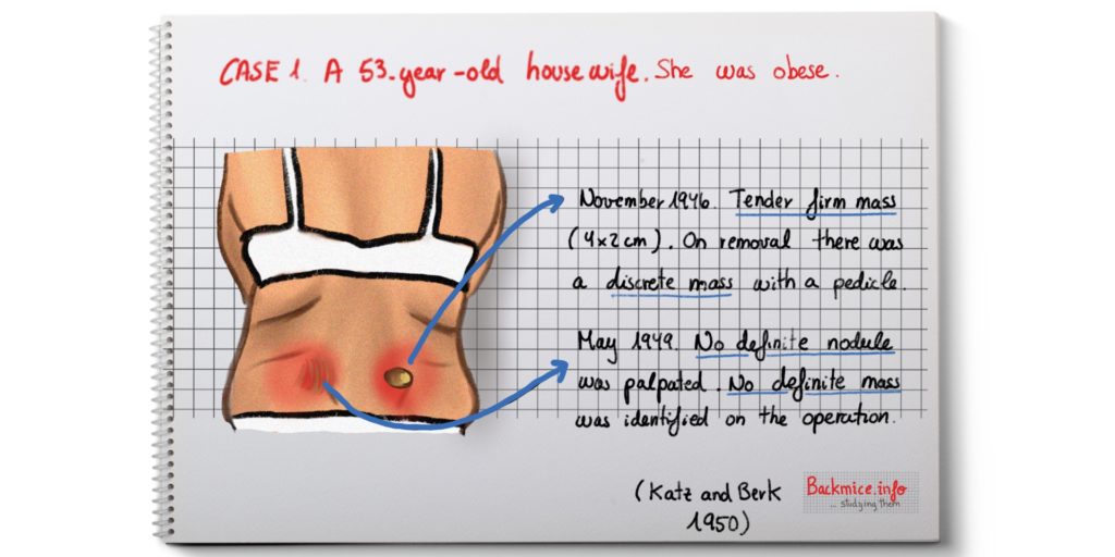
A 53-year-old housewife that was examined in 1946. She complained of a dull pain over the right lumbar area. She explained a relatively sudden onset and that she had been with pain for 2 weeks.
Then she recalled that 2 years before she had had a similar pain on the left side that lasted for 1 week and cleared spontaneously.
The patient was OBESE, weighting 189 pounds. Examination of the body was negative. But on the right sacroiliac articulation, deeply placed, a tender, firm mass, measuring 4 by 2 cm could be palpated.
Local infiltration with 2% procaine solution DID NOT diminish the pain.
She was admitted in hospital. They operated on her, and found a fatty mass with a PEDICLE deep through the fascia. The pedicle was cut, the mass removed and the fascia repaired. Afterwards, the pain promptly disappeared.
Histological examination showed NORMAL FATTY tissue.
The patient was seen again 3 years later presenting an identical pain in the left lumbar area. The pain was worse while she was upright and better if she was lying down. The examination showed a tender subcutaneous mass overlying the sacroiliac joint in a similar position as the previous one. This mass was less well defined. They COULD NOT find a definitive mass, instead they just remove some of the fibrofatty tissue, and then there was complete relief. She kept out of pain at least for 6 years.
DISCUSSION
They mention previous works:
– In 1935 Sutro reported the high incidence of subcutaneous nodes in the sacroiliac region; they were present in 94 of the 170 unselected hospital patients. And 33 from 94 presented low back pain at the moment of examination. Despite he removed fat nodules in 4 patients and 1 got free from pain, Sutro didn’t appreciate the full significance of these fatty tumors as cause of low back pain.
-Two years later, RIES gave more significance to the epsiacroiliac lipomas cases, he explored 1,000 patients, of whom 1/3 had lumbosacral backache, and of that 1/2 were found to have subcutaneous lumbosacral tumors. An equal number had asymptomatic nodules in the same location. He removed some of the lipomas surgically and it showed to be lipomas with a FIBROUS CAPSULE. He called these nodules EPISACROILIAC LIPOMAS.
-In the British literature, these nodules became to be designated “fibrositis”.
–Copeman and Ackerman removed these nodules in 10 British soldiers. In all cases there was relief from symptoms. Then they did further studies in cadavers and find out that even in cachectic bodies there was what they called BASIC FAT PADS. They correlated the distribution of the basic fat pads with the presence of the “trigger points” known in the “fibrositis”.
In a later paper, they noticed that in some areas the fibro-fatty tissue may become EDEMATOUS, because of the fibrous investment, the painful TENSION may then be set up. If the tension persisted, there could also be herniation of the lobules through the fascia.
–Herz (1945 and 1946) ALSO did anatomical studies in cadavers and agreed with the BASIC FAT PATTERN of Coperman and Ackerman. Herz mentioned that in OBESE persons this basic fat pad could become obscured by a more generalized deposition of fat. Herz also mentioned that the fascias were NOT of uniform thickness, being thinner in some places than in others, being frequent the presence of deficiencies of the fascial membranes. In these places, the underlying fat tended to BULGE through, occasionally resulting in complete herniation. These may not give any symptom until a trauma or lying in bed for several days may produce edema and perpetuation of the condition. He also related the low back pain with obese women that seemed to present a thinning of the fascia membrane. Sometimes, in obese women it was not possible to dissert a definite mass that would correspond with a palpable nodule, anyway they would do a wide dissection of fat.
Their results: 5 cases of obese women with episacroiliac lipomas
The authors present a number of patients with low back pain that presented OBESITY. They relate that obese women have higher risk of presenting this kind of pathology.

About case number 3: She weighed 110 pounds at the time of marriage, soon after 2 years she weighed 235 pounds. She stated that her lower back pain started when she gained weight. After surgical excision of the mass, she experienced prompt relief. THE LOCAL ANESTHETIC injection DID NOT give relief to the patient.
Published in July 2018 By Marta Cañis Parera 
References
Sutro CJ. Subcutaneous fatty nodes in the sacroiliac area. Am J Med Sci 1935; 190:833-837.
Reis E. Episacroiliac lipoma. Am J Obstet Gynecol 1937;34:492-8.
Copeman, W.S.C. and Ackerman, W. Fibrositis of the back (1944) . Quart.J.Med. 13,37.
COPEMAN WS, ACKERMAN WL. Fatty herniation in low back pain. Lancet. 1947 Aug 2;2(6466):188. PubMed PMID: 20255787.
R HERZ. HERNIATION OF FASCIAL FAT AS A CAUSE OF LOW BACK PAIN WITH RELIEF BY SURGERY IN SIX CASES. 1945;128(13):921–925. doi:10.1001/jama.1945.02860300011003
HERZ R. Herniation of subfascial fat as a cause of low back pain; report of 37 cases treated surgically. Ann Rheum Dis. 1946 Dec;5(6):201-3. PubMed PMID: 20242353.
HENCH PS. Discussion of the paper by Ralph Herz: herniation of subfascial fat as a cause of low back pain. Ann Rheum Dis. 1946 Dec;5(6):204. PubMed PMID: 20242354

