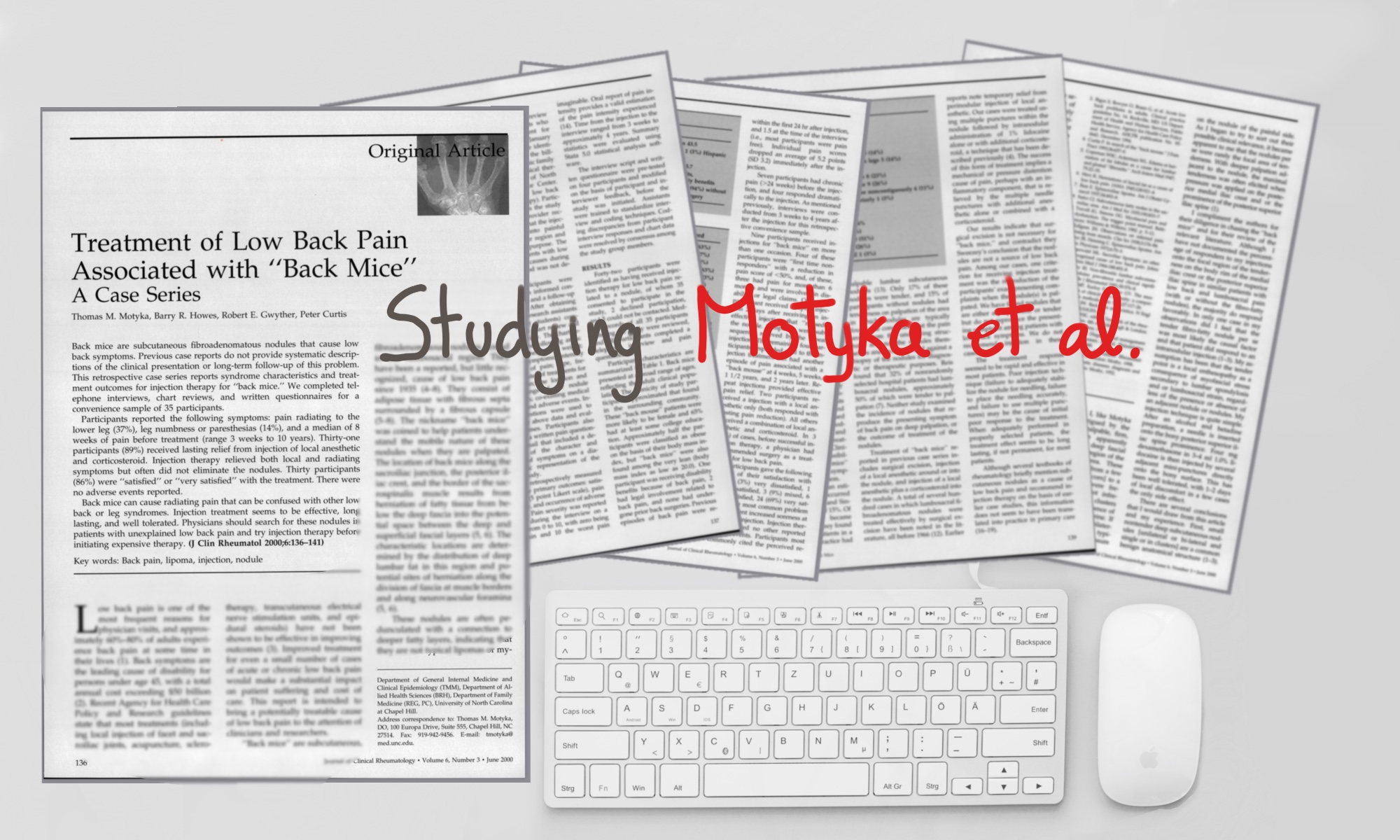
This article about a cause of nonspecific low back pain was published in the Journal of Clinical Rheumatology in June 2000 (the last author is Peter Curtis, who already had previous publications about back mice).
Motyka et al. present a RETROSPECTIVE case series STUDY about the treatment with local anesthetic to patients with nonspecific low back pain syndrome associated with back mice.
Previous studies didn’t present a long-term follow-up of the treatment of this entity. That’s why they presented this report that they completed by telephone interviews, chart reviews and questionnaires with a sample of 35 participants.
-31 participants (89%) received LASTING RELIEF from injection of local anesthetic and corticosteroid. That relieved both the local and the radiating symptoms but mainly didn’t eliminate the nodules.
-30 participants (86%) were “satisfied” o “very satisfied” with the treatment. There were no adverse events reported.
Notes on the article:
Treatment of Low Back Pain associated with “back mice”
Motyka TM, Howes BR, Gwyther RE, Curtis P.
From the Department of General Internal Medicine and Clinical Epidemiology.
J Clin Rheumatol. 2000 Jun;6(3):136-41.
The low back pain problem
The authors start commenting the high prevalence of nonspecific low back pain and its cost and the fact that there aren’t effective treatments. So a successful treatment even if it were for a small number of cases, would make a SUBSTANTIAL IMPACT with patient suffering and cost care.
The authors’ main aim with this paper is: “To intend to bring a POTENTIALLY TREATABLE CAUSE of nonspecific low back pain to the attention of clinicians and researchers that is underdiagnosed”.

What are “back mice” according to Motyka et al.?
“Back mice” are subcutaneous fibroadenomatous nodules found in the lumbosacral region. They have been reported since 1937 by Ries, but had received little recognition.
They consist of ADIPOSE TISSUE with fibrous septa surrounded by a FIBROUS CAPSULE.
The nickname “back mice” was coined to help patients understand the mobile nature of these nodules when they were palpated.
They are mainly located along the sacroiliac junction, the posterior iliac crest and the border of the sacrospinalis muscle.
They are the result from HERNIATION of fatty tissue from below the deep fascia into the potential space between the DEEP and SUPERFICIAL FASCIA layers. Especially, the herniation can occur along the muscle borders and the neurovascular foramina.
The nodules are OFTEN PEDUNCULATED with a connection to deeper fatty layers, indicating that they aren’t typical lipomas or the known as MIOFASCIAL trigger points.
Back mice can be differentiated from trigger points, since miofascial trigger points are FLAT and lie within the muscle. Back mice are SUPERFICIAL to muscle tissue.
The mechanism producing the LOCAL and REFERRED symptoms of “back mice” is not clear. It has been related to the compression to the neurovascular bundle. It can have a sclerotomal path that may confuse the clinicians with “nerve root compression”.
Back mice had been reported to be treated with: local injection, dry needling or surgical excision.
ATTENTION: Motyka et al. warns that back mice can mimic vertebral disk disease, so failure to recognise this entity may lead to unnecessary testing and inappropriate treatments. They also warn that clinicians are generally UNAWARE of the existence of back mice.

They did the follow up of 35 patients.
Methods
They collect RETROSPECTIVE data from patients with NONSPECIFIC LOW BACK PAIN that received local injections in the tender nodules from the period of 1994 to 1998.
Results
-The back mouse patients were 65% women.
-HALF of the participants were obese according to their body mass index.
-Most participants had a SINGLE painful nodule.
-Many pieces of information like the number of nodules, the size, etc. could not be collected.
-Symptoms associated to the nodules include numbness and paresthesias distant from the nodule. Pain could radiate to the knee.
Reaction after local injection:
-PAIN (from 1-10): from 7.6 to 2.4 within the first 24 h. At the time of study it was 1.5, since most of the participants were pain-free.
-4 patients were “first time non responders”, one of these got better after a second injection (first missed “the spot”).
-Most of them received injection with local anesthetic and corticosteroid, two patients just with local anesthetic.
-IN 3 CASES before the successful injection treatment, the patients had been suggested surgery by another physician to treat the low back pain.
-68% of the patients gave a satisfying rating of VERY SATISFIED.
-The only common problem as adverse event reported by patient was TRANSIENT INCREASED SORENESS at the site of injection.
DISCUSSION about NONSPECIFIC LOW BACK PAIN and BACK MICE
-Most cases of low back pain are attributed by physicians as NONSPECIFIC low back pain, once serious causes such fracture, infection or herniated intervertebral disc are ruled out.
-They presented a study that shows that some cases of nonspecific low back pain are associated to painful lumbar adenomatouse nodules that can be easily treated by injection therapy.
-Prevalence of these nodules varies in different studies. Some are asymptomatic and other tender or painful.
-The injection technique with multiple punctures suggests that the cause of pain is related to mechanical or pressure distention. Some techniques inject directly the nodule and others perinodularly.
-Their results contradict the results of Swezey that suggested that the pain arises from deeper structures than the nodules themselves.
-Poor response to the injection technique could be: failure to adequately stabilize the nodule for needling, failure to place the needling accurately, and failure to MULTIPLE punctures.
-When the technique is placed in properly selected patients, the treatment seems to be long lasting, if not permanent, for most patients.
-Several textbooks of rheumatology (like Copeman 1969) briefly mention the SUBCUTANEOUS NODULES as a cause of low back pain and recommend injection therapy, too; this information DOES NOT SEEM TO HAVE BEEN TRANSLATED INTO PRACTICE in primary care.
-The authors admit that the study has certain bias, like the fact of being a retrospective study without group control. Nevertheless, the immediate dramatic improvements in the patients suggest that the treatment is effective.
THE COMMENTARY OF Robert L. Swezey
From the Arthritis & Back pain center. Medical director, California.
Dr. Swezey starts the commentary saying that 10 years before, he was also intrigued by the presence of small palpable, firm, movable, and often apparently tender deep subcutaneous nodules. The size from these nodules ranged from a few mm like a kernel of corn to cm like a grape. They are frequently BILATERAL and sometimes occurred in clusters overlying the bony prominence of the posterior bony prominence of the superior iliac crest.
If the nodules were found bilateral, tenderness would typically be noted only by pressure on one side.
He was intrigued and started to study them. He noticed that with deeper palpation adjacent to the nodule there was elicited pain. So he hypothesized that the nodules ARE NOT the MAIN CAUSE of pain, they just happened to be there. He injects local anesthetic with or without the presence of the nodules because he thinks the problem is a focal enthesopathy secondary to underlying lumbar spondylosis or lumbosacral strain.
He injects 4 mg of dexamethasone in 3-4 ml of 1% lidocaine. He performs mini-punctures in the bony surface of the posterior superior iliac crest. There can be local discomfort for 1-2 days as a side effect.
Swezey says that regarding Motyka’s article several considerations had to be done:
- He says that according to his experience the NON-TENDER SUBCUTANEOUS NODULES (unilateral, bilateral, single or in clusters) are a common BENIGN ANATOMICAL STRUCTURE.
- These nodules correspond physiological herniation of the deep layer of fatty tissue and may occasionally become incarcerated and undergo a painful inflammatory process.
- But Swezey also injects even with the absence of the nodulations, that’s why he thinks that the problem is a lumbosacral enthesopathy.
- He hypothesizes that in the cases that the doctors just inject the nodule; it may give relief by the diffusion from the nodule to the adjacent enthesopathic iliac crest.
Published in June 2018 by Marta Cañis Parera 
References
- Motyka TM, Howes BR, Gwyther RE, Curtis P. Treatment of low back pain associated with ;;back mice”: a case series. J Clin Rheumatol. 2000 Jun;6(3):136-41. PubMed PMID: 19078461
- Curtis P. In search of the ‘back mouse’. J Fam Pract. 1993 Jun;36(6):657-9. PubMed PMID: 8505609.
- COPEMAN WS, ACKERMAN WL. Fatty herniation in low back pain. Lancet. 1947 Aug 2;2(6466):188. PubMed PMID: 20255787.
- HERZ R. Subfascial fat herniation as a cause of low back pain; differential diagnosis and incidence in 302 cases of backache. Ann Rheum Dis. 1952 Mar;11(1):30-5. PubMed PMID: 14915430; PubMed Central PMCID:PMC1030570.
- Reis E. Episacroiliac lipoma. Am J Obstet Gynecol 1937;34:492-8.
- Sutro CJ. Subcutaneous fatty nodes in the sacroiliac area. Am J Med Sci 1935; 190:833-837.
- Pace JB, Henning C. Episacroiliac lipoma. Am Fam Physician. 1972 Sep;6(3):70-3. PubMed PMID: 4262540.Singewald ML. Another cause of low back pain: lipomata in the sacroiliac region. Trans Am Clin Climatol Assoc. 1966;77:73-9. PubMed PMID: 4223124; PubMed Central PMCID: PMC2441105
- Swezey Non-fibrositic lumbar subcutaneous nodules: prevalence and clinical significance. Br J Rheumatol. 1991 Oct;30(5):376-8. PubMed PMID: 1833023.


